Abstract: The study of pathogenesis of many diseases, including cancer metastasis, requires quantification of cell-cell and cell-extracellular matrix interactions on the tissue level. A particularly promising assay technology is high throughput multiphoton tissue cytometry. This technique is based on mulitphoton microscopy (MPM) allowing minimally invasive 3D resolved imaging deep inside tissues. MPM not only can provide tissue and cellular morphological information but can also provide genetic (via fluorescence in situ hybridization), proteomic (via antibody labeling) and biochemical (via fluorescence spectroscopy with appropriate biochemical probes) information. A major limitation of microscopy is its relatively low speed and only a small tissue volume can be assayed greatly limiting statistical power of the assay. We have developed 3D tissue cytometry based on high speed MPM imaging by producing and simultaneously scanning multiple two-photon excitation foci simultaneously. This approach parallelizes the raster scanning process by acquiring data over multiple regions in the specimen. The advantage of this method is that the imaging speed is enhanced by the degree of parallelization but the pixel dwell time can in principle remain the same as in the conventional multiphoton microscope preserving image signal to noise ratio. To overcome the depth penetration limitation of TPM in studying thicker specimens, we integrated an automated, motorized, microtome into a high-speed TPM system. By alternating and overlapping optical sectioning with mechanical sectioning, it is possible to rapidly image samples with arbitrary thickness. Whole organ imaging with subcellular resolution will be demonstrated.
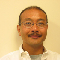
Biography: Peter So is a professor in the Department of Mechanical and Biological Engineering in the Massachusetts Institute of Technology. Prior to joining MIT, Peter So obtained his Ph.D. from Princeton University in 1992 and subsequently worked as a postdoctoral associate in the Laboratory for Fluorescence Dynamics in the University of Illinois in Urban-Champaign. His research focuses on developing high resolution and high information content microscopic imaging instruments. These instruments are applied in biomedical studies such as the non-invasive optical biopsy of cancer, the mechanotransduction processes in cardiovascular diseases, and the effects of neuronal remodeling on memory plasticity.

